Neurovascular Signalling Research Group
Neurovascular Signalling research group
- Department of Biomedicine, Aarhus University
One of the primary interest of our research group is neurovascular coupling, the fine-tuning signalling that enables an adequate, specially restricted blood supply in accordance to metabolic demand of neuronal tissue. Disturbances in neurovascular coupling have detrimental consequences for brain integrity and neuronal function, as it seen in stroke, chronic stress, major depression and migraine. Our laboratory aims to uncover the key molecular mechanisms behind neurovascular coupling, and highlight their significance in pathologies.
The group is working in a close proximity with several groups in DANDRITE, CFIN, as well as with Cardiovascular Phenotyping Core Facility at the Department of Biomedicine. We are focused on in-vivo Laser Speckle Contrast brain imaging on anaesthetized and awake mouse models. Changes in neurovascular responses to sensory stimulations and after stroke are assessed. These further studied in brain slices (parenchymal arteriole diameter and intracellular Ca2+ changes in response to electric field stimulation) and in isolated blood vessels ex-vivo. The molecular background is studied by proteomics and spatial transcriptomics.
Research focus
- Neurovascular coupling signalling
- Cerebral blood flow regulation; the role of ion transport, e.g., the Na+, K+-ATPase, Cl- and K+ channels
- Stroke, futile reperfusion
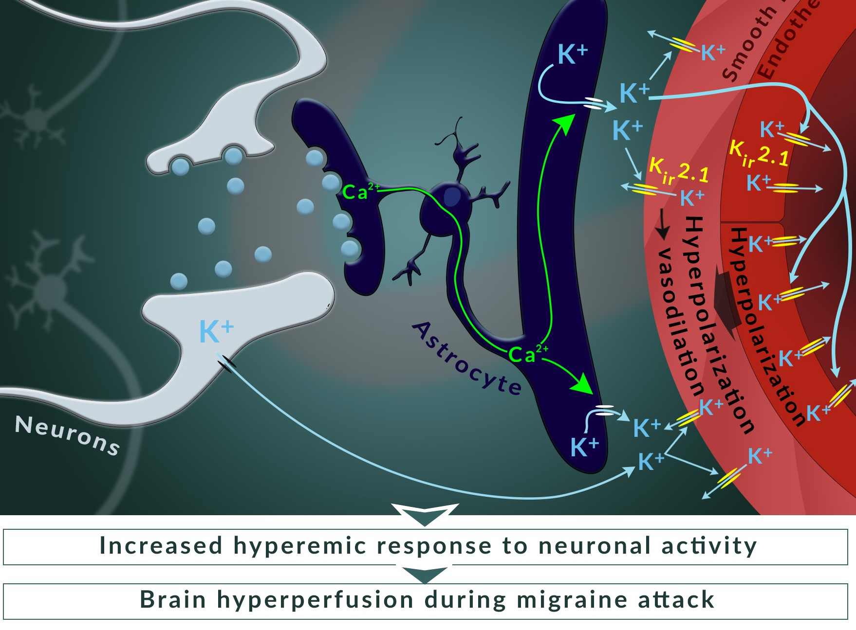
- Cerebrovascular abnormalities in inherited migraine
- Cerebrovascular abnormalities in major depression and chronic stress
- Co-morbidity of metabolic disturbances and cerebrovascular dysfunctions
Techniques
- Laser Speckle Contrast Imaging in awake and anaesthetized rodents
- Brain slices to assess parenchymal arteriole diameter and intracellular Ca2+ responses to neuronal excitation
- Telemetry: blood pressure, blood glucose, biopotentials (ECG, EEG), activity and body temperature
- Blood pressure, blood flow measurements in anaesthetized and awake mice
- Glucose/insulin tolerance test
- Ischemic stroke rodent models
- Patch clamp, sharp electrode membrane potential measurements
- Small artery myography
- Intracellular Ca2+, H+ imaging with ion selective dyes
- Protein and PCR lab, spatial transcriptomics
Contact
PI/Centre leader/head of centre/main contact person:

Illustration:
Staehr C., Rajanathan R., Postnov D.D., Hangaard L., Bouzinova E.V., Lykke-Hartmann K., Bach F.W., Sandow S.L., Aalkjaer C., Matchkov V.V. Abnormal neurovascular coupling as a cause of excess cerebral vasodilation in familial migraine. Cardiovasc Res. 2020;116(12):2009-2020. doi: 10.1093/cvr/cvz306.